Attempto! 02/2023: Seeing through tumors
Immunotherapies use T cells to target and attack tumors. With state-of-the-art imaging, cancer researcher Bettina Weigelin can even watch this happening in real time.
The PET scan shows a cross-section of a man’s body with shadows in the lungs, abdomen, arms and legs: cancer metastases. “Here you can see a stage IV melanoma,” Bettina Weigelin explains the image on a large monitor at the Werner Siemens Imaging Center (WSIC) at the University Hospital Tübingen. In the next image, taken a few weeks later, almost all the shadows have disappeared, except for three tumors in the arm and leg.

An impressive result for immunotherapy that has unleashed the body’s own defense against cancer within twelve weeks. However, the chances are still not good for the patient: “These three metastases are very likely to lead to death,” is the sobering prognosis. “It’s not enough to catch 99 percent of the tumor cells. We have to get them all.”
Molecular cell biologist Bettina Weigelin is Professor of Preclinical Imaging of the Immune System at the Faculty of Medicine and belongs to the only oncological cluster of excellence in Germany: “Image-Guided and Functionally Instructed Tumor Therapies (iFIT)”, where scientists are researching new, individualized cancer therapies.
In her group at the WSIC, Weigelin is working towards finding out why immunotherapies often do not work adequately. She is investigating immune reactions against tumors using a state-of-the-art intravital microscope with optical lenses connected in series, mirrors, high-resolution objectives, and three high-energy infrared lasers.
Researchers can use the astounding device to observe what is happening in living tissue both in cell cultures and in mice in real time – which is why it is called “intravital”.
It’s not enough to catch 99 percent of the tumor cells. We have to get them all.
Behind the scenes
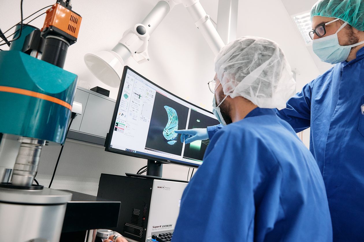
One floor lower into the laboratory area of the WSIC, we put on a protective suit, hood and mask and pass through an airlock. Since 2021, this space houses the intravital microscope system which costs around one million euros. With the lasers, it covers several square meters.
Weigelin uses controllers to adjust the laser light to the correct wavelength. It can show individual cells in tissue labeled with fluorescent proteins. Scientists can look up to two millimeters deep into the tissue using the microscope. “We are not limited to the surface which is the advantage over other microscopy methods,” she explains.
Although images of the body can also be produced with Magnetic Resonance Imaging (MRI) or Positron Emission Tomography (PET) – this method uses radioactive tracers to examine metabolic processes or track specific cells in the body – there are limits to both methods when it comes to resolution. “We can detect the tumor, but not its individual cells which we need to investigate the efficacy of immunotherapies which happen at the cellular level. Intravital microscopy closes a resolution gap in imaging.”
Promising immunotherapy
Since 2011, impressive advances have been achieved by immunotherapies. Checkpoint inhibitors enable the immune system to attack tumors. “This does not work for every patient, long-term tumor remission is currently only achieved in a few patients,” says Weigelin.
Also with cell-based immunotherapies, Weigelin sees room for improvement. To treat cancer using T-cell therapy, T cells are first harvested from the patients’ blood. They have the task of recognizing and destroying harmful cells, such as tumor cells. Once those T cells have been modified with genetic methods or modified viruses to identify cancer cells, they are injected back into the body.
The difficulty with immunotherapies is that tumor cells are the body’s own cells. Normally, complex mechanisms prevent the immune system from targeting the body’s own cells. Eliminating these mechanisms works relatively well for some types of cancer. “In studies of advanced melanoma, immunotherapy has allowed about 20 percent of patients to live at least ten years longer. The response rate, where cancer growth was transiently decreased, was even higher. But there are types of cancer where it doesn’t work at all.”
In the tumor, there are many strategies that our body uses to prevent an immune reaction against the body’s own cells. We need to recognize and understand every hurdle.
Immune cells battle in real time
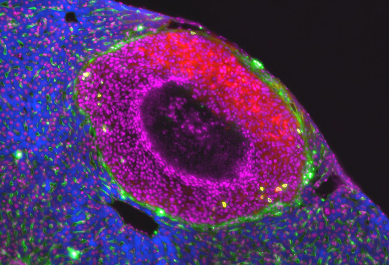
Weigelin shows us a film from the intravital microscope. We watch how lively green spots – the T cells – target tumors which are shown in red until the tumor disintegrates into small pieces. Once the enemy is destroyed, the T cells swim to the next victim. “In this cell culture, therapeutically modified T cells are destroying melanoma cells,” she explains. “The T cells recognize certain molecules on the tumor surface and then effectively kill the cancer cells.”
Another film shows how T cells surround individual tumor cells – from a melanoma in the skin of a mouse. “The T cells were altered to recognize tumor cells efficiently, and then injected into the mouse. From there, they were able to target certain molecules in the tumor as intended. But it’s not enough to kill the tumor completely.”
Modifying the T cells is only half the battle, as Weigelin says. A crucial point is understanding the tumor microenvironment. In the tumor, there are many strategies that our body uses to prevent an immune reaction against the body’s own cells. “We need to recognize and understand every hurdle. This requires different strategies.” When she zooms in, we can see that more T cells dock in individual areas of the mouse tumor than in others. “Laser microscopy allows us to make detailed observations at the cellular level.”
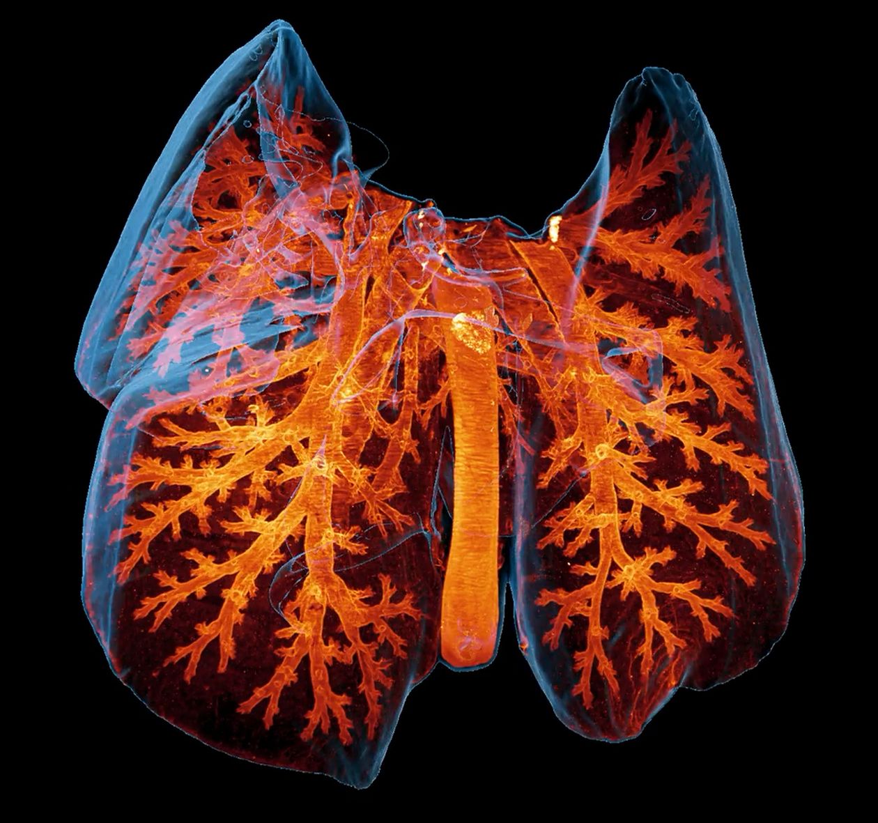
Transparent lungs
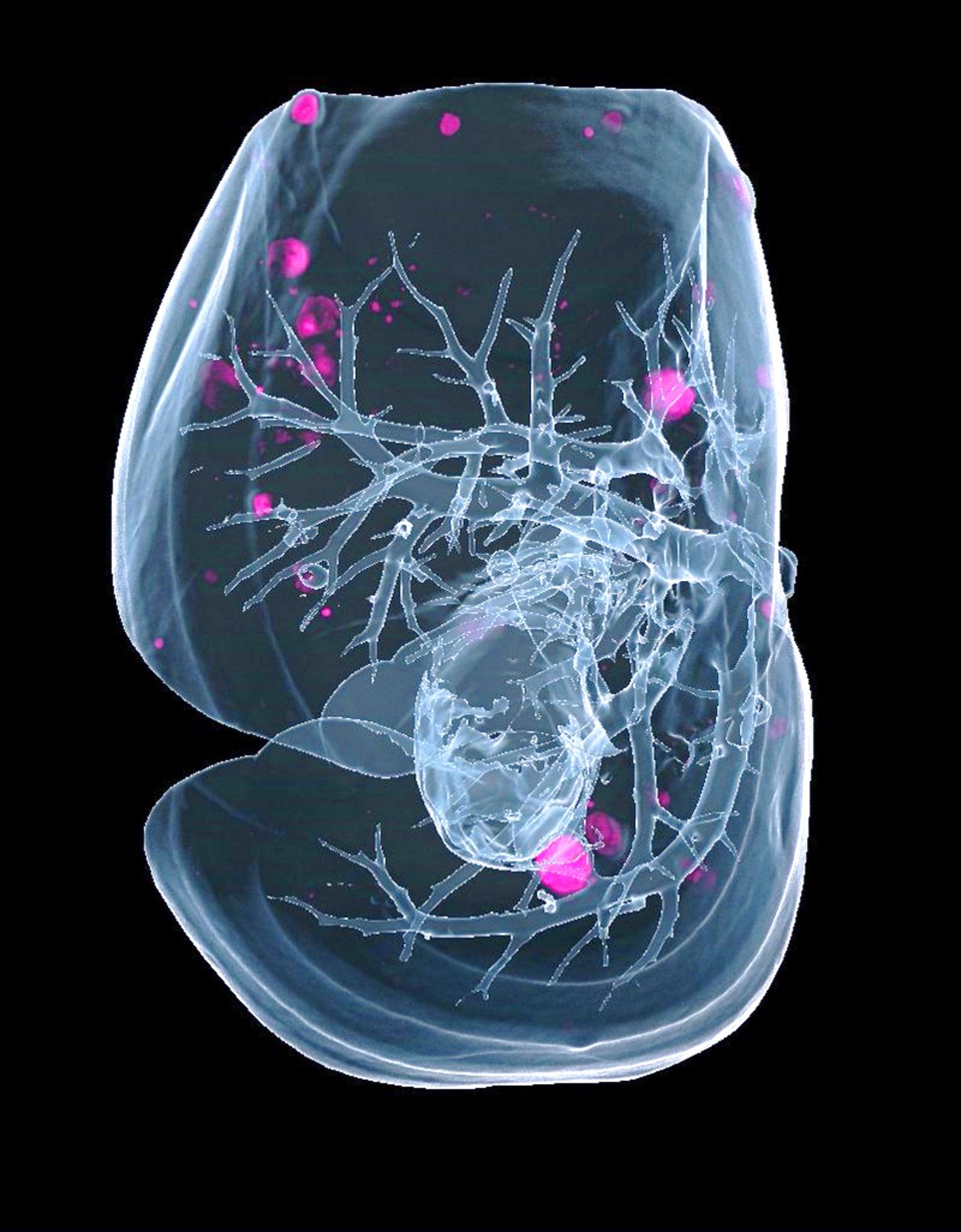
Four years ago, Weigelin’s team established another method for imaging at the WSIC: light sheet microscopy. It is used to investigate the development and treatment options for metastases. “Metastases are often hidden deep inside of an organ,” she explains. “It would make things easier if we could make the organ transparent. Then we could use a microscope to show us the tumor cells there.”
In light sheet microscopy, large pieces of tissue are first made transparent by dissolving out the light-reflecting lipids, dyes and proteins by using chemicals. Weigelin zooms in on an image of a mouse lung until an isolated tumor cell lights up. “These cells have survived the therapy and are apparently resistant. They are often responsible for the cancer coming back.”
Light sheet microscopy makes these hidden cells visible. Scientists can use models – such as mice – to test how immunotherapies affect individual tumor cells. “We can use this method to learn which therapies could be combined. We might observe that with a certain therapy, individual tumor cells disappear in the lungs, but survive in the liver. With other therapies, it may be the other way around.”
In addition to cancer, microscopy methods are also suitable for research into other diseases, such as inflammation or autoimmune diseases. Imaging has the opposite interest here: The immune response should not be unleashed but suppressed.
In basic research, our goal today shoud be, to cure cancer.
“We are no longer completely in the dark in the fight against cancer,” summarizes Weigelin. “My feeling is that we have understood enough to improve existing therapies and develop new ones.“ She doesn’t want to be content with keeping the cancer at bay for a few months. “At least in basic research, our goal today should be to find out how we can cure cancer.”
Text: Christoph Karcher